Cyclone Separator,Pump Lquid Separator,Pump Hydrocyclone Separators,Pump Liquid Separation Dandong Huarui Fluid Machinery Co., Ltd , https://www.ddhrseal.com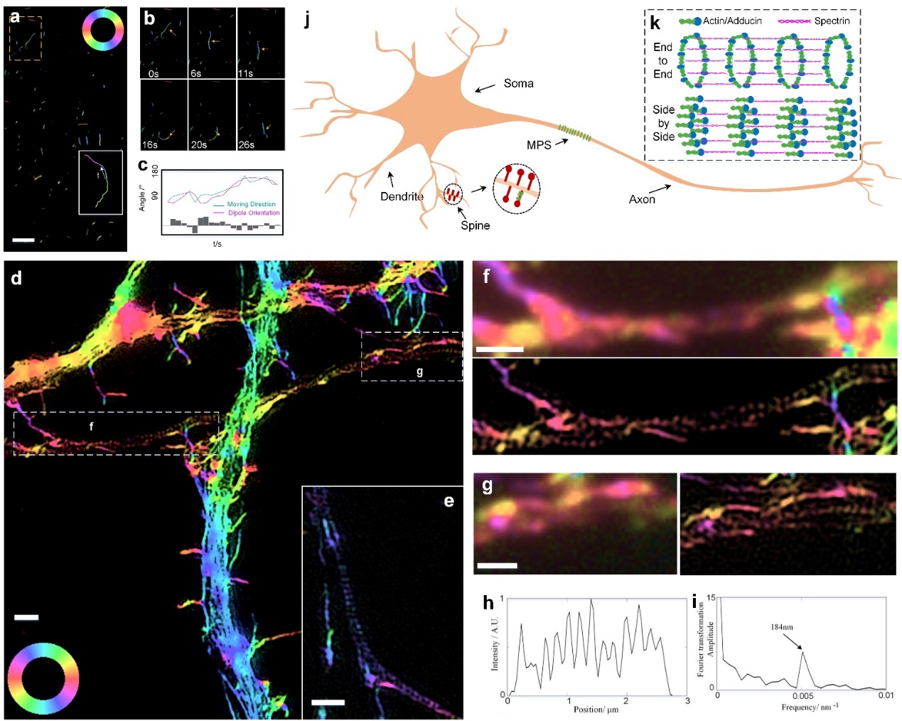
Peking University Institute of Technology has made important progress in super-resolution microscopy imaging
[ Instrument Network Instrument Development ] Polarization is one of the basic physical properties of electromagnetic waves. Polarization characteristics have been widely used in the fields of light field control, microscopic imaging, quantum optics, and stereoscopic display. In biology, the orientation of a target protein can be revealed by measuring the dipole orientation of the fluorophore by polarization imaging. Although the super-resolution microscopy technology can break through the diffraction limit of light and achieve high-resolution imaging at 100 nm scale, it is extremely limited in application due to the inability to know the orientation of biomolecules.
In order to study the localization and orientation of proteins in subcellular structures, the research group and research colleagues of Peking University of Engineering, Peking University recently developed polarized light structured light microscopy (pSIM). The related research results were published in the journal Nature Communications on October 16th under the title "Super-resolution imaging of fluorescent dipoles via polarized structured illumination microscopy".
Structured Light Imaging (SIM) is highly compatible with bio-imaging because of its high resolution and fast imaging speed, which is favored by biologists. Drawing on the principle of SIM imaging, Xi Peng et al. constructed a space-azimuth high-dimensional composite space, and extracted the azimuth and spatial super-resolution information of the fluorescent dipole, thus realizing polarization-structured light imaging.
In order to verify the wide compatibility of this technology with SIM, the researchers tested a variety of commercial SIM systems and self-built SIM platform, as well as 2D-SIM, 3D-SIM, TIRF-SIM imaging capabilities, successfully extracted the dipole of fluorescent molecules. Sub-azimuth information and super-resolution structure information. They conducted extensive biological experiments to demonstrate their broad applicability, such as the interaction between actin filament, actin and myosin in λ-DNA, BAPE cells and mouse kidney tissue, and GFP-stained U2OS live cell microtubules. The team studied the membrane-associated cycle skeleton (MPS) in neurons. With high spatial resolution and accurate polarization detection, pSIM reveals a new model of "side-by-side" assembly of actin rings in MPS, overturning the "end-to-end" structural hypothesis of actin rings in previous textbooks. With high spatiotemporal resolution and unique dipole orientation information, pSIM has broad application prospects in solving various biological problems in the future.
pSIM reveals a new model of "side by side" assembly of actin rings in MPS
Generally speaking, an innovative technology usually adopts the following two ways to benefit the scientific research community: 1. Open access to relevant technologies, other scholars can obtain applications by building similar systems; 2. Commercialize relevant technologies, and other scholars purchase instruments. To get the app. This work opened up a third way to promote scientific research: by digging deep into the potential characteristics of SIM technology and commercial instruments, the existing SIM system was “powered†and excavated existing SIMs that were not noticed by the inventors. The inherent polarization detection characteristics of the system enable the existing system to implement the function of the polarization SIM without any modification. This allows many life science laboratories with existing SIM systems to perform direct polarization SIM analysis, which will greatly advance the research of polarization super-resolution imaging.
In recent years, Xi Peng's research group has been working on the development of polarization super-resolution technology and SIM super-resolution technology, such as: 1. Fluorescence dipole super-resolution technology (SDOM) using polarization characteristics, published in Light: Science and Applications, and obtained Nature Methods. Highly rated (related links http://news.pku.edu.cn/xwzh/129-295505.htm); 2, SDOM applied to gold nanoparticles SERS super-resolution imaging (Nanoscale 2018); 3, developed Reduced frame SIM technology to increase the rate of structured light imaging by more than 2 times (IEEE TIP2018); 4. Participated in the development of Hessian-SIM ultra-high-speed structured light imaging technology, and proposed rolling SIM technology, which can improve SIM imaging speed by more than 3 times ( Nature Biotechnology 2018). These scientific advances have laid a solid foundation for this work.
Xi Peng and Academician Dai Qionghai from Tsinghua University are co-authors of this work. Dr. Zhang Wei, the first author and co-author of the joint communication, received the post-doctoral program of Peking University, and the first author Chen Xingyu is a doctoral student in the Department of Automation of Tsinghua University. The nerve cell experiment in this paper was completed in cooperation with the research group of Professor Zhang Yan of the McGovern Brain Science Center of Peking University. The in vitro actin experiment was completed by Professor Li Xiangdong of the Institute of Zoology of the Chinese Academy of Sciences. The live cell microtubule imaging was performed by Peking University School of Life Sciences. Chen Xiaowei's research team completed the cooperation. The SIM super-resolution microscopy of this work was completed on the biomicroscopic platform of Peking University, and was helped by teachers such as Shan Chunyan. The work was funded by the National Natural Science Foundation of China, the Ministry of Science and Technology, the Outstanding Youth Science Fund of the Beijing Municipal Science and Technology Commission, and the Clinical +X Project and Instrument Specialization of Peking University.